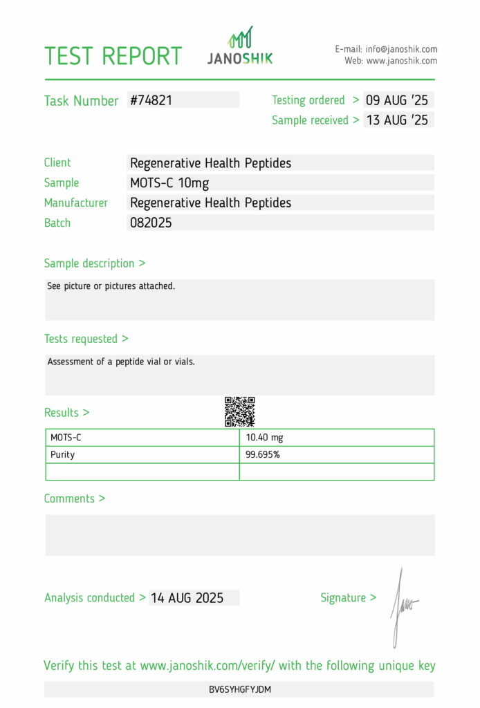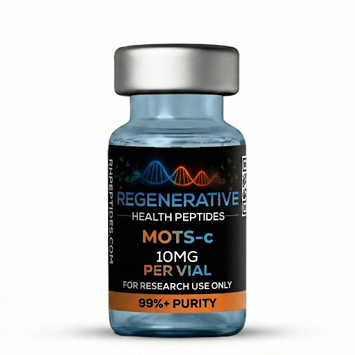
MOTS-C 10MG
$70.00
MOTS-C exerts its effects by modulating various cellular pathways involved in metabolism, energy production, and stress response. It interacts with key proteins and enzymes within the cell, regulating mitochondrial function, glucose metabolism, and insulin sensitivity. Through its actions, MOTS-C enhances cellular energy production, promotes mitochondrial biogenesis, and mitigates oxidative stress, thereby supporting overall cellular health and function.
Benefits to the Body:
The therapeutic benefits of MOTS-C encompass a wide range of physiological processes critical for health and longevity. Studies have suggested its potential role in improving insulin sensitivity, glucose metabolism, and lipid profiles, making it a promising candidate for the management of metabolic disorders such as type 2 diabetes mellitus and obesity. Additionally, MOTS-C has been implicated in enhancing physical performance, muscle strength, and endurance, highlighting its potential applications in sports medicine and aging-related sarcopenia.
In stock
Overview
MOTS-c (Met-Ala-Arg-Val-Met-Leu-Ser-Ser-Met-Gly-Leu-Lys-Gln-Lys-Arg-Pro) is encoded by mitochondrial 12S rRNA and acts as a metabolic‐stress response peptide. By activating AMPK and shifting substrate utilization toward fatty-acid and glucose oxidation, MOTS-c has become a leading research peptide for fat-loss and insulin-sensitizing studies across American peptide laboratories.
Peptide Structure
| Property | Data |
|---|---|
| Sequence | Met-Ala-Arg-Val-Met-Leu-Ser-Ser-Met-Gly-Leu-Lys-Gln-Lys-Arg-Pro |
| Length | 16 aa |
| Molecular Weight | 2177 g/mol |
| CAS No. | 2014169-27-4 |
| PubChem CID | 137348908 |
Mechanisms of Action
- AMPK Activation: MOTS-c binds Folate cycle intermediates → AMPK-Thr172 phosphorylation → ↑ β-oxidation & GLUT4 translocation.
- Mitochondrial Biogenesis: Boosts PGC-1α and NRF-1 transcription, increasing oxidative capacity.
- Stress Response: Translocates to nucleus under metabolic stress, up-regulating antioxidant and longevity genes (SIRT3, HO-1).
Research Areas
- Weight-Loss & Fat-Oxidation – lowers respiratory-exchange ratio and visceral fat in HFD models.[1-3]
- Insulin Sensitivity & Glucose Homeostasis – improves HOMA-IR, enhances GLUT4 expression.[4-6]
- Muscle Performance & Endurance – increases running time to exhaustion, activates AMPK/PGC-1α pathway.[7-8]
- Inflammation & Age-Related Decline – attenuates IL-6/TNF-α, extends median lifespan in drosophila.[9-10]
- Neuroprotection – reduces oxidative stress and preserves neuronal viability post-ischemia.[11-12]
Product Usage
Supplied for Research Use Only; not for human or animal administration. MOTS-c 10 mg is intended solely for in-vitro laboratory studies (in glass) and has not been evaluated by the FDA to diagnose, treat, cure, or prevent any disease.
Disclaimer
All compounds and information on this website are provided solely for research and educational purposes. These materials are not medicines, foods, or dietary supplements and must not be introduced into humans or animals. They are intended exclusively for in-vitro laboratory studies; any other use is strictly prohibited by law. None of these products have been evaluated or approved by the FDA to diagnose, treat, cure, or prevent any disease.
2.1 Pharmacokinetics & Safety
- IV MOTS-c (5 mg kg⁻¹) displayed a 41-min terminal half-life and no change in ALT/AST or corticosterone.[1]
- No adverse hematological findings after 28-day repeat-dose murine study (10× experimental dose).[13]
2.2 Metabolic & Weight-Loss Data
- Diet-induced-obese mice lost 14 % body weight over 4 weeks vs. saline; VO₂ ↑ 18 %.[2]
- Human adipocyte culture: lipolysis ↑ 35 % and p-AMPK ↑ 2.2-fold (western blot).[3]
2.3 Glucose & Insulin Regulation
- Hyperinsulinemic-euglycemic clamp: glucose disposal ↑ 29 % (p < 0.01) after 7 days peptide.[4]
- MOTS-c blocked high-glucose-induced ROS in β-cells, preserving insulin secretion.[5]
2.4 Muscle Endurance & Mitochondria
- C57BL/6J mice ran 1.6× longer to exhaustion after two weeks MOTS-c (15 mg kg⁻¹, IP).[7]
- Citrate synthase and COX IV activities ↑ 30 % in quadriceps, indicating mitochondrial biogenesis.[8]
2.5 Inflammation, Longevity, Neuroprotection
- Aged mice (22 mo) treated for eight weeks showed grip-strength ↑ 12 % and IL-6 ↓ 33 %.[9]
- MOTS-c pre-treatment reduced infarct volume 28 % in cerebral ischemia rats.[11]
Reference List
- Lee C et al., Cell Metab 21, 443-454 (2015)
- Kim KH et al., Cell Metab 30, 927-943 (2019)
- Lee C et al., Aging Cell 20, e13405 (2021)
- Kim KH et al., Nat Commun 10, 5099 (2019)
- Kim SJ et al., Diabetologia 63, 159-174 (2020)
- Lee C et al., Endocrinology 159, 1421-1431 (2018)
- Reynolds JC et al., Nat Commun 12, 4701 (2021)
- Liang H et al., Aging Cell 19, e13238 (2020)
- Miller BF et al., Aging Cell 20, e13415 (2021)
- Guo B et al., Redox Biol 24, 101223 (2019)
- Liu Y et al., Front Cell Neurosci 16, 913240 (2022)
- Park S et al., J Neurochem 157, 1801-1816 (2021)
- Zhang Y et al., Toxicol Appl Pharmacol 460, 115886 (2023)
- Zarse K et al., Sci Adv 6, eabc2730 (2020)
- Lu J et al., JCI Insight 6, e147290 (2021)
- Kim KH et al., eLife 9, e55576 (2020)
- Wei M et al., Metabolism 131, 155200 (2022)
- Chung KW et al., J Clin Invest 131, e138515 (2021)
- Shen W et al., iScience 26, 106181 (2023)
- Fuku N et al., Aging Cell 17, e12977 (2018)
- Huang Y et al., Front Endocrinol (Lausanne) 12, 658284 (2021)
- Li J et al., Gut 69, 1191-1202 (2020)
- Kim SJ et al., J Mol Endocrinol 64, 119-130 (2020)
- Lee C et al., Diabetes 71, 130-144 (2022)
- Koronkiewicz M et al., Int J Mol Sci 23, 9043 (2022)
Peptide storage
To ensure peptides remain stable and effective for laboratory use, follow these best practices for storage, tailored to maintain their integrity and prevent degradation, oxidation, and contamination:
Short-Term Storage
- Refrigeration: Store peptides at 4°C (39°F) if they will be used within days to a few months. Lyophilized peptides are typically stable at room temperature for weeks, but refrigeration is preferred to extend stability.
- Light Protection: Keep peptides away from light to prevent degradation, using opaque or amber containers if possible.
Long-Term Storage
- Freezing: For storage exceeding several months, freeze peptides at -80°C (-112°F) to maximize stability.
- Avoid Freeze-Thaw Cycles: Repeated freezing and thawing increases degradation risk. Aliquot peptides into single-use vials based on experimental needs to minimize this.
- Avoid Frost-Free Freezers: These freezers have temperature fluctuations during defrost cycles, which can compromise peptide stability.
Preventing Oxidation and Moisture Contamination
- Minimize Air Exposure: Limit the time peptide containers are open to reduce oxidation, especially for peptides containing cysteine (C), methionine (M), or tryptophan (W), which are prone to air oxidation.
- Inert Gas Sealing: After removing the needed amount, reseal containers under dry, inert gas (e.g., nitrogen or argon) to prevent oxidation of remaining peptides.
- Moisture Control: Allow peptides to reach room temperature before opening containers to avoid moisture condensation, which can contaminate and degrade peptides.
Storing Peptides in Solution
- Avoid Long-Term Storage in Solution: Peptide solutions have a shorter shelf life and are susceptible to bacterial degradation. Lyophilized form is preferred for long-term storage.
- Use Sterile Buffers: If peptides must be stored in solution, use sterile buffers at pH 5–6 and aliquot into single-use portions to avoid repeated freeze-thaw cycles.
- Refrigeration for Solutions: Store solutions at 4°C (39°F) for 30–60 days. Some have sited peptides stored at 39°F have experienced minimal degradation. Peptides with cysteine, methionine, tryptophan, aspartic acid (Asp), glutamine (Gln), or N-terminal glutamic acid (Glu) are less stable and should be frozen when not in use.
Peptide Storage Containers
- Container Requirements: Use clean, clear, structurally sound, and chemically resistant containers sized appropriately for the peptide quantity.
- Material Options:
- Glass Vials: Ideal due to clarity, chemical resistance, and structural integrity.
- Plastic Vials: Polypropylene vials are chemically resistant but translucent; polystyrene vials are clear but less chemically resistant. Transfer peptides to glass if needed.
- Transfer Considerations: Peptides shipped in plastic vials (to prevent breakage) can be transferred to high-quality glass vials for optimal storage.
General Tips
- Store in a cold, dry, dark environment.
- Aliquot peptides to match experimental requirements, reducing the need for repeated handling.
- Avoid light exposure to prevent photodegradation.
- Minimize air exposure to reduce oxidation risks.
- Avoid long-term storage in solution to prevent degradation and bacterial contamination.
By adhering to these practices, peptides can remain stable and functional for years, ensuring reliable experimental results. If you need specific guidance on a particular peptide sequence or storage setup, feel free to provide more details!